
We bring the latest technology together with a skilled,
caring team to produce accurate diagnoses in a timely manner.
1100 Monterey Street
Suite 210
San Luis Obispo, CA 93401
(805) 542-9700
Hours: Monday to Friday 8 a.m. – 5 p.m.
Details & Directions: San Luis Diagnostic Center
Find us on Facebook
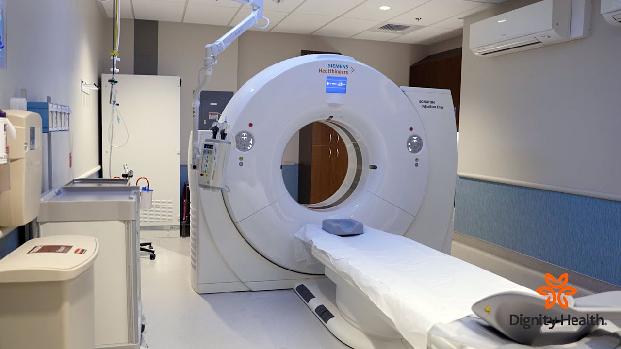
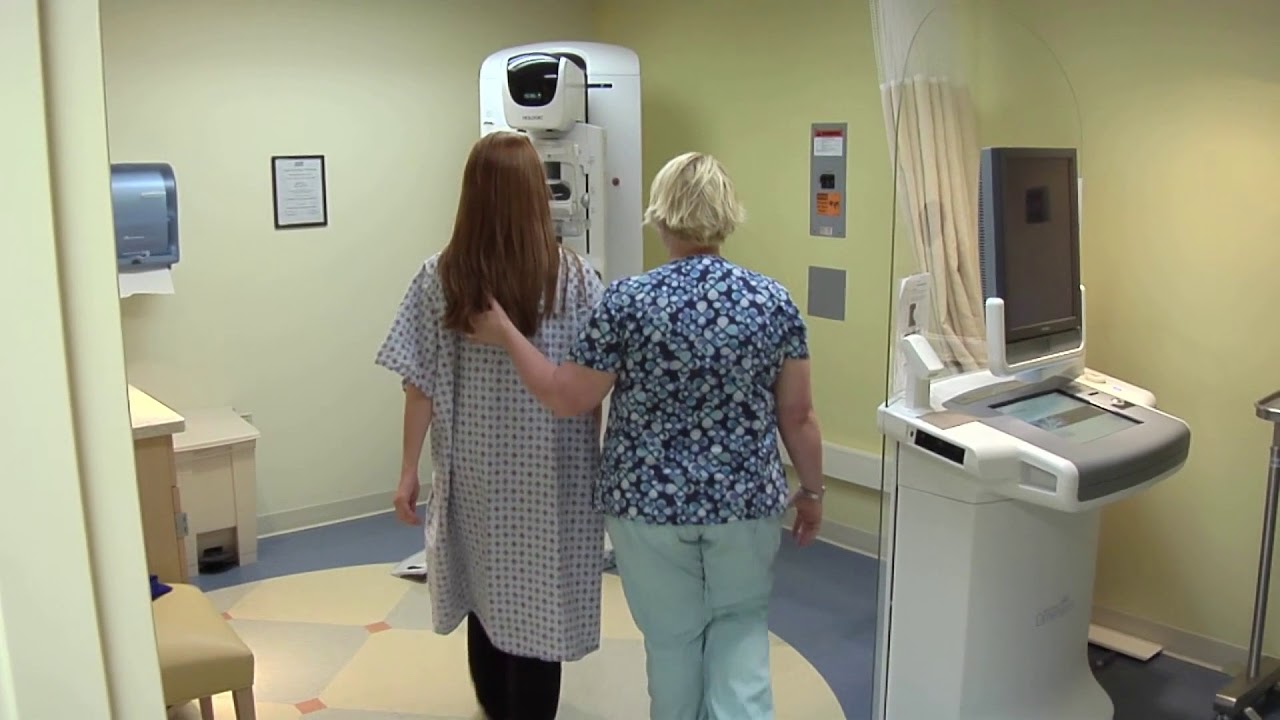
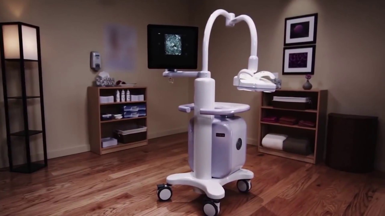
San Luis Diagnostic Center is a full-service diagnostic facility. We have the most advanced equipment available and offer a number of diagnostic imaging services:
San Luis Diagnostic Center is home to some of the most advanced imaging technology available. These state-of-the-art tools mean we produce the high quality, detailed images physicians need to make an accurate diagnosis to determine the correct treatment for you. And we can do it faster than any other facility in the area.
Not only can the advanced equipment we use complete examinations quickly, reducing the stress or anxiety that can sometimes accompany imaging studies, but our radiologists interpret studies and have reports to your physician by the next day. In addition, our sophisticated digital imaging system means that images can be viewed online by a physician.
Our center was designed with our patients in mind. The unique nature of our facility lead GE to select San Luis Diagnostic Center as a GE Show Site. This distinction means that GE brings potential customers from all over the world to observe our facility and GE equipment at work. We have educated physicians from as far away as China and India.
San Luis Diagnostic Center is part of French Hospital Medical Center therefore we contract with most major insurance companies and bill patients’ insurance companies for them. Prior to an examination, we contact all patients for details regarding their insurance coverage. Any amounts not covered by insurances, such as deductibles or co-insurance amounts, we collect at the time of service. For our patients’ convenience, we accept personal checks and credit cards.
Specific insurance inquiries are handled by our business office at (805) 542-9700.
San Luis Diagnostic Center is regularly featured on HealthBreak, Dignity Health’s health education program on KSBY.
Featuring Fred Vernacchia, MD, Medical Director, San Luis Diagnostic Center.
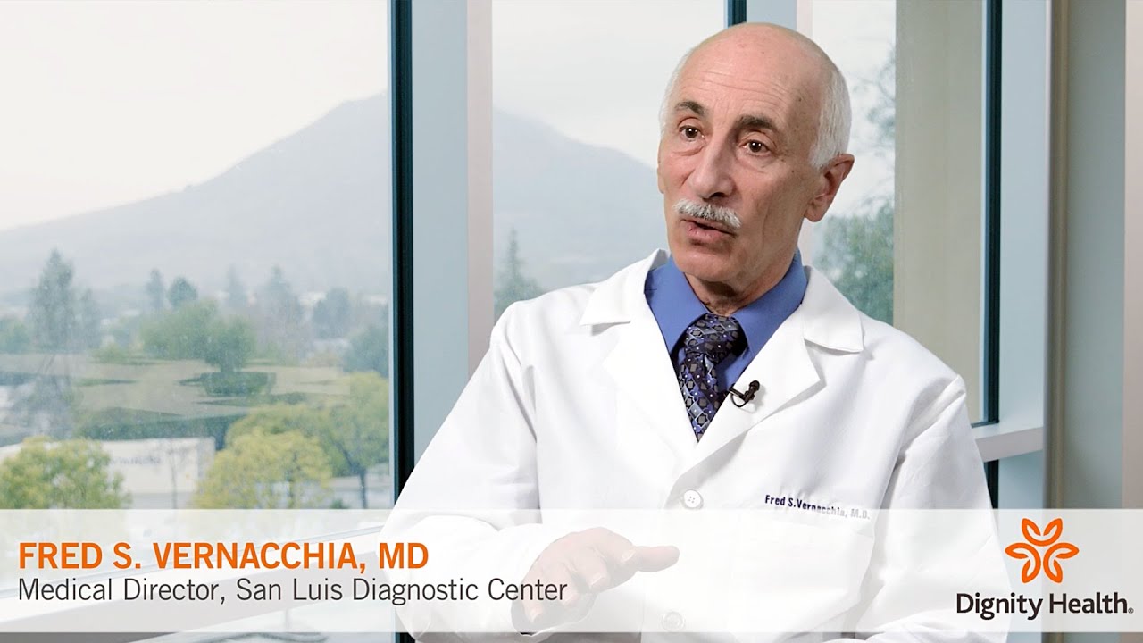
Featuring Fred Vernacchia, MD, Medical Director, San Luis Diagnostic Center.
Featuring Fred Vernacchia, MD, Medical Director, San Luis Diagnostic Center.
Featuring Fred Vernacchia, MD, Medical Director, San Luis Diagnostic Center.
Featuring Fred Vernacchia, MD, Medical Director, San Luis Diagnostic Center.
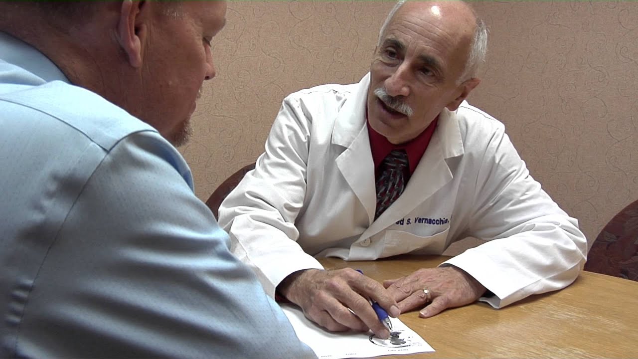
Featuring Fred Vernacchia, MD, Medical Director, San Luis Diagnostic Center.
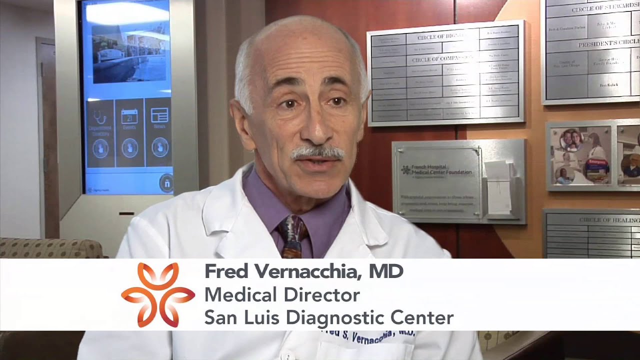
Featuring Fred Vernacchia, MD, Medical Director, San Luis Diagnostic Center.

My Care – Dignity Health is convenient, secure, electronic way to access your medical records information. All you need is internet access and an e-mail address.
Features of the Patient Portal
Get started, it’s as easy as 1-2-3!
Download the My Care by Dignity Health app for convenient access on the go. Text MY CARE to (602) 600-0605 to get the install link or visit the app store today!
![]()
![]()
Physicians can access the San Luis Diagnostic Center (PACS) images and reports by using our eJacket Mobile Viewer or McKesson Radiology Station Lite desktop software. Please see the following documents for additional information: